Hyperparathyroidism – primary, secondary, tertiary – simplified

The disease of hyperparathyroidism is diverse.  The causes of it are very different.  As a simplification there’s primary, secondary and tertiary hyperparathyroidism:

All three of these involves increased PTH (parathyroid hormone)

Primary: elevated calcium, low phosphorus in blood stream.  Usually caused by a parathyroid adenoma.

Secondary: normal or low calcium, usually caused by renal failure.

Tertiary: failure of treatment of secondary hyperparathyroidism to correct increased PTH.

More details on hyperparathyroidism

Primary hyperparathyroidism is most commonly caused by adenomas, greater than 85%.  Next most common cause would be hyperplasia which is 10% of the causes, and there is a 1% possibility that primary hyperparathyroidism is caused by a carcinoma.

Risk factor for primary hyperparathyroidism includes family history, MEN I and MEN IIa, and also radiation.  Major difference between MEN IIA and IIB:  MEN IIA has Hyperparathyroidism – hyperplasia, MEN IIB does not.  More on MEN syndromes.

Sestamibi scan is a method of detecting hyperparathyroidism.

Carcinomas which are commonly associated with hypercalcemia:

  • Breast Cancer Metastasis
  • Postate Cancer
  • Kidney Cancer
  • Lung Cancer

Another cause of hypercalcemia:  FHH – Familial hypocalciuric hypercalcemia

  • autosomal dominant
  • asymptomatic hypercalcemia and low urine calcium
  • with or without elevated PTH
  • No treatment is generally required for FHH
 

Primary hyperaldosteronism

Primary hyperaldosteronism

  • Hypertension in the presence of elevated aldosterone and suppressed plasma renin
  • Two major causes are aldosteronoma and idiopathic hyperaldosteronism (IHA)

Aldosteronoma

  • 60% of cases
  • more often in women
  • Hypertension and hypokalemia is more severe compared with IHA
  • Salt loading does not change aldosterone levels
  • CT usually reveals a small solitary adrenal tumor
  • Adrenal vein sampling will reveal unilateral hypersecretion of aldosterone.
  • Treatment is surgical resection

Idiopathic Hyperaldosteronism (IHA)

  • 30% of cases
  • More often in Men
  • Salt loading decreases aldosterone levels
  • CT reveals normal to bilaterally enlarged glads
  • Adrenal vein sampling reveals bilateral hypersecretion of aldosterone
  • Treatment is with potassium sparing diuretics – spironolactone


Somatostatinoma, Glucagonoma, insulinoma, vipoma, gastrinoma

Pancreas NeoEndocrine/Islet cell Tumors

Sometimes associated with MEN I (multiple endocrine neoplasia)

Somatostatinoma

  • tumor of the delta cells of the endocrine pancreas
  • triad of: Diabetes Mellitus, Steatorrhea, and Gallstones
  • Most are malignant and have metastasized

Glucagonoma

  • Necrolytic Migratory Erythema (NME).  found in 70%
  • Diabetes Mellitus
  • Most are malignant and have metastasized

VIPOMA

  • Vasoactive Intestinal Polypeptide tumor
  • Verner Morrison syndrome
  • Causes WDHHA:  Watery Diarrhea, Hypokalemia, Hypochlorhydria, Achlorhydria
  • Activation of cellular adenylate cyclase and cAMP production
  • 50% malignant

Insulinoma

  • 10% malignant
  • Whipple’s triad of pancreatic insulinoma – Hypoglycemia, symptoms of hypoglycemia, relief of symptoms when glucose is given.

Gastrinoma

  • Zollinger-Ellison Syndrome
  • Excess gastric acid production
  • Can arise in the duodenum or pancreas
  • 60% malignant


What is the RET proto-oncogene?

RET Proto-Oncogene

From an insurance standpoint on RET proto-oncogene testing for MTC – found on the internet:

Aetna considers diagnostic testing for germline point mutations in the RET gene medically necessary for members with apparently sporadic medullary thyroid carcinoma.

What is a proto-oncogene?

A proto-oncogene is a normal gene which can become an oncogene due to mutations or increased expression.

Inflammatory Bowel Disease – Topics in General Surgery

Inflammatory Bowel Disease = Crohn’s disease and Ulcerative Colitis.

Crohn’s Disease

  • Mouth to anus
  • Skip areas
  • Full thickness
  • Anal involvement common (fistulas, abscess, fissures, ulcers)
  • Cancer risk less than that of ulcerative colitis

Ulcerative Colitis

  • Bloody diarrhea
  • Colon only
  • Anal involvement is rare
  • Always involves the rectum and spreads proximally (no skips)
  • mucosa, submucosa (not full thickness)
  • 20% risk of colon cancer after 20 years of disease


Normal Lab Values – Topics in General Surgery

To help remember these numbers I’ve simplified the ranges.  Please look up these actual values in other sources, these numbers are just easier to remember without a written reference.  These are just crude numbers within the normal range.  The units have also been left off.  So this really is not a reliable source of info.

Amylase = 100

BUN (Urea Nitrogen) = 15

Bilirubin Total = 1

Calcium = 10

Creatinine = 1

Sodium = 140

Chloride = 100

Potassium = 4

Bicarbonate = 25

Magnesium = 2

WBC = 10

Phosphorus = 4

Upper GI Bleeding – Topics in General Surgery

Upper GI (gastrointestinal) Bleeding

  • bleeding proximal to the ligament of Trietz
  • Most common cause of UGI bleeding is Duodenal Ulcer (25%), Gastric ulcer (20%), Acute Gastritis (15%)
  • EGD is the diagnostic test of choice with UGI bleeding

Peptic Ulcer Disease

  • Both gastric and duodenal ulcers

Duodenal Ulcers

  • Caused by increased gastric acid production
  • ZE syndrome (zollinger ellison) is a risk factor

Surgeries for duodenal ulcers

  • Graham patch
  • Truncal Vagotomy and pyloroplasty
  • Truncal Vagotomy and antrectomy with Billroth I (one limb off the stomach remnant)
  • Truncal Vagotomy and antrectomy with Billroth II (2 limbs off stomach remnant)
  • Proximal gastric vagotomy (Highly selective)
  • Truncal Vagotomy – removal of a segment of vagal trunk on the distal esophagus, this decreases gastric acid secretion
  • Drainage procedure is needed with Truncal Vagotomy

Gastric Ulcers

  • Due to decreased cytoprotection, gastric acid is usually normal or low.
  • Most of  the gastric ulcers (70%) are on the lesser curvature.  5% is on the greater curvature.
  • With all gastric ulcers, must rule out gastric cancer  – need biopsy

Mallory Weiss Syndrome

  • Postemesis longitudinal tear
  • Do not use sengstaken-blakemore balloon

Esophageal Variceal Bleeding

  • Need to verify bleeding with EGD.  Only 50% of UGI bleeding in patients with known Esophageal Varices are bleeding from varices.
  • Treatment:  sengstaken-blakemore balloon, Liver transplant, shunts (ie. Warren)

Lower GI Bleeding – Topics in General Surgery

Lower GI Bleeding

  • Distal to ligament of Treitz
  • Most common causes of lower GI bleeding:  1. Diverticulosis (usually right sided), 2. Vascular ectasia
  • Must rule out upper GI bleeding with lower GI bleeding.  Use NGT aspiration and/or EGD.
  • If massive bleeding – consider angiogram
  • If slow bleeding – consider colonoscopy
  • If colonoscopy is negative consider tagged RBC study
  • tagged RBC scan (radiolabeled) can detect bleeding at a rate of 0.1 ml/Min
  • Angiogram detects 1ml/Min


Tall Cell Variant of Papillary Thyroid Cancer

Prognosis for the tall cell variant of papillary carcinoma

Tall cell variant of papillary thyroid cancer has been shown to be more aggressive and have a worse prognosis than the typical papillary thyroid cancer.

See more info regarding papillary thyroid cancer and other thyroid cancers.

Favorable characteristics for papillary thyroid cancer

  • age less than 50 for women
  • age less than 40 for men
  • tumor less than 5 cm.

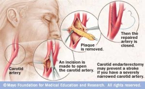
Indications for a Carotid Endarterectomy

When is a Carotid Endarterectomy Performed

Asymptomatic patients:

  • A Carotid Endarterectomy is indicated in patients with stenosis greater than 60%

Symptomatic patients:

  • A Carotid Endarterectomy is indicated in symptomatic patients with stenosis greater than 70%
  • If there are multiple episodes of TIAs, then operate when the stenosis is greater than 50%

carotidendarterectomy

Picture credit:

http://www.mayoclinic.com/health/medical/IM01657

Disclaimer: These are notes only.  Every patient and condition is different and requires a in person consultation with a medical doctor.  No medical advice is contained here, just academic discussion which may be very different from real life.   There’s always ongoing trials and the information here may be out of date.

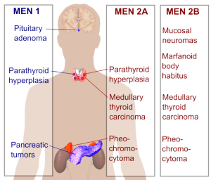
Multiple Endocrine Neoplasias

MEN syndromes

Inherited as autosomal dominant

All family members of a patient diagnosed with MEN syndromes should be screened for MEN.

Types:  MEN I, MEN IIA, MEN IIB

MEN I

Most common tumors:  mnemonic – PPP (all the P’s are followed by a vowel)

Other tumors associated with MEN I = adenomas of Adrenal 30% and Thyroid 15%

MEN IIA

Most common tumors: mnemonic MPH – 2 miles per hour

  • Medullary thyroid carcinoma 100%
  • Pheochromocytoma 33%
  • Hyperparathyroidism (50%), hypercalcemia

100% of patients with MEN IIA have medullary carcinoma of the thyroid.

MEN IIB

Most common abnormalities: mnemonic MMMP – 3M plastics

  • Mucosal neuromas 100%
  • Medullary thyroid carcinoma 85%
  • Marfanoid
  • Pheochromocytoma 50%

Physical findings of MEN IIB:  Mucosal neuromas (mouth, eyes, etc), Marfanoid body shape, abnormal arch of foot, constipation.

Major difference between MEN IIA and IIB:  MEN IIA has Hyperparathyroidism – hyperplasia, MEN IIB does not.

MEN I and MEN IIA have hyperplasia of the parathyroid – treated with removal of all parathyroid tissue with autotransplant of some parathyroid tissue – perhaps into the forearm.

Multiple_endocrine_neoplasia

Picture credit:

http://commons.wikimedia.org/wiki/User:Mikael_H%C3%A4ggstr%C3%B6m


Types of Thyroid Cancer

Five main types of Thyroid Cancer

  1. Papillary Carcinoma (80%)
  2. Follicular Carcinoma (10%)
  3. Medullary Carcinoma (5%)
  4. Hurthle Cell Carcinoma (4%)
  5. Anaplastic/Undifferentiated Carcinoma (1-2%)

P’s of Papillary Thyroid Cancer:

  • Popular (most common type – 80%)
  • Psammoma bodies
  • Palpable Lymph Nodes – common
  • Positive Iodine 131 uptake
  • Positive Prognosis
  • Postoperative Iodine 131 to diagnose / treat metastasis if a total thyroidectomy was done.

F’s of Follicular Cancer of the Thyroid

  • Far away metastasis (spreads hematogenously)
  • FNA NOT (FNA cannot be done to diagnosis Follicular Cancer of the thyroid
  • Female predominance (3:1)

M’s of Medullary Cancer of the Thyroid

  • MEN II (Multiple Endocrine Neoplasia type II)
  • aMyloid
  • Median Lymph Node dissection
  • Modified neck dissection if lateral nodes are positive
  • Secretes Calcitonin
  • Stimulated by Pentagastrin
  • Cancer of the Thyroid C cells = parafollicular cells

Calcitonin (tumor marker for MTC)

  • Reduces blood levels of Calcium
  • Opposed the effects of PTH (parathyroid hormone)
  • Calcitonin is used as a tumor marker for medullary thyroid cancer (MTC)

Pentagastrin

  • Pentagastrin has effects like gastrin
  • Pentagastrin is used as a stimulation test to elevate serotonin levels and cause symptoms of carcinoid syndrome, provoking flushing.
  • Pentagastrin has been used to stimulate ectopic gastric mucosa for the detection of Meckels diverticulum by nuclear medicine.
  • Pentagastrin-stimulated calcitonin test is a diagnostic test for MTC (Medullary carcinoma of the thyroid).  In patients with suspected MTC but borderline levels of calcitonin, injecting pentagastrin will cause calcitonin levels to rise tremendously.


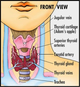
Thyroid Anatomy
Thyroid Anatomy

Picture from: http://www.washingtonendocrineclinic.com/Thyroid-Care.html


Neck Anatomical Zones for Cancer and Trauma

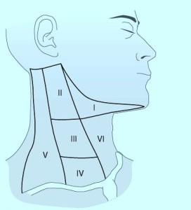
Neck Zones for Cancer

When thinking of neck zones, there are those zones used in trauma and there are those used for cancer surgery.

The neck zones or levels depicted here are for cancer surgery.  Lymph nodes or neck masses in these areas are described using these locations:

anatomical zones of neck
Picture copied from internet

  • In patients presenting with solid neck masses – those masses associated with the thyroid move with swallowing.  Those in levels 3,4, and 5 do not move with swallowing.
  • Metastatic lymph nodes found in level 6 – could come from thyroid.
  • U/S and FNA should be performed after exam/history for Solid Neck Mass.
  • With well differentiated thyroid cancers, there is a high rate of cervical metastasis particularly with papillary cancers.
  • With lateral lymph nodes involved in thyroid cancer, survival rate is not improved significantly with LN dissection in the neck for patients older than 60 years old or for men with a 4 cm or larger well differentiated cancer.
  • Prophylactic lymph node dissections does not seem to improve survival with patients undergoing thyroid cancer treatment.
  • If thyroid tumor is small with documented (U/S) lateral lymph nodes.  There is a benefit to dissecting level 3 or 4 lymph nodes (lymph node dissection or modified dissection) in the younger patient.

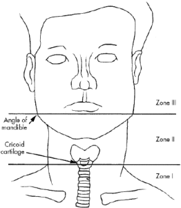
Trauma neck zones

picture copied from internet:

neck.zones


Legal and Ethics for Surgeons – Highlights – Notes

Legal Issues and Ethics

  • When a patient has a DNR order and needs a surgery with anesthesia, consider discussing the revoking the DNR order temporarily or focusing on what the patient wants for quality of life.
  • Unrelated urgent surgery to terminal illness + DNR order = some hospitals rescind the DNR order x 24 hours with careful documentation of the time.
  • High quality X-ray and multiple views of the X-ray may be required when looking for a possible retained sponge from surgery.
  • Some surgeons believe that the patient should be informed when the sponge / instrument counts are inaccurate at the end of a case even if there is no retained objects found on X-ray.
  • Laws vary regarding protection from apologizing.
  • Negligence = failure to exercise standard of care (reasonably prudent person / same situation).
  • First element of informed consent, decide whether the person is competent of making decisions on behave of themselves, their child or their family member.
  • To honor a patient’s request to make a medical/surgical decision for them, you must have already established a relationship with the patient, no other good options, believe that the course of the option is best for the patient.
  • Parents disagreeing, first determine if only one parent or both parents have the legal right to give permission.
  • A durable power of attorney is a type of advanced directive which may include other legal issues such as financial issues.
  • The advanced directive of a patient trumps the wishes or demands of a family member or surrogate.
  • A surgeon’s fine motor skills doesn’t appear to decline with age.


Anesthesia and Pain Management for the Surgeon – Highlights – Notes

Anesthesia and Pain Management

  • PRIS – Propofol Infusion Syndrome – can be fatal.  Cardiac failure, rhabdomyolysis, and a severe metabolic acidosis.
  • Adrenal insufficiency secondary from Etomidate (induction agent) administration may be a cause of hypotension in the post op surgical patient.
  • Dexmedetomidine  (Precedex)has  analgesic properties, sedation, stable respiratory rates, predictable cardiovascular responses.
  • Ativan should not be discountinued abruptly after long term use in the ICU patient.
  • Heterotopic calcification and myopathies are complications of long term chemical paralysis – thus chemical paralysis for ventilated patients are now not as popular.
  • Pulse oximetry readings not as accurate in patients with smoke inhalation.
  • Colorimetric CO2 indicator – stomach intubation can still change the color, initially.
  • CNS and Cardiotoxity related to local anesthetic use – a concern when intercostal nerve blocks used for multiple rib fractures.  Lidocaine info
  • Place/Pull epidural catheters, won’t do unless lovenox/heparin sq held for 24 h.
  • As doses and duration of Metaclopramide (Reglan – Dopaminergic blocker) increases – neurologic side effects (movement disorders) also increases.
  • Oral to IV morphine is 3:1
  • Main use for LMA is for short cases in operating room.  There are concerns for vomiting and aspiration.


Precedex does not cause respiratory depression

Dexmedetomidine (Precedex)

hp-precedexLogo

  • Sedation without respiratory depression
  • Ideal for use in the Intensive Care Unit.
  • Can be used for managing extubations.
  • No absolute contraindications
  • Compared to Versed (Midazolam), Precedex is associated with less delirium, tachycardia, and hypotension, but had more bradycardia
  • Agonist of alpha 2 adrenergic receptors in the brain
  • FDA reports that infusions greater than 24 hours in duration include ARDS, respiratory failure, and agitation


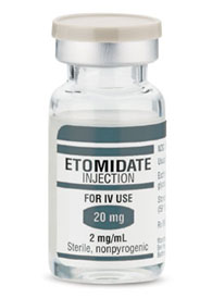
Etomidate – drug for anesthetic induction

Etomidate

Etomidate Vial

Background info

  • Imidazole
  • Short acting IV anesthetic
  • Hypnotic effects
  • Amnestic properties, no analgesic properties
  • works through the GABA adrenergic system
  • Used often in Rapid Sequence Intubation
  • Short duration of action, rapid onset
  • Excellent hemodynamic profile
  • Cerebroprotective effects including reduction of intracranial pressure.  Used with patients with traumatic brain injury.
  • Etomidate has hemodynamic stability which is better than all other induction agents except possibly Ketamine.
  • Post operative vomiting is more common with Etomidate than with other induction agents.
  • Requires a slow push for intubation induction otherwise one will get trismus
  • Onset of action is 30-60 seconds.

Etomidate and Adrenal Insufficiency

  • Adrenal supression:  inhibits cortisol production of the adrenal gland
  • Etomidate reversibly inhibits 11-beta-hydroxylase
  • Etomidate is no longer used for continuous sedation

Signs and Symptoms of Adrenal Insufficiency

  • Hypotension
  • Orthostatic Hypotension
  • Weakness
  • Dizziness
  • Hypoglycemia
  • Dehydration
  • Weightloss
  • Nausea
  • Vomiting
  • Diarrhea
  • Cardiovascular collapse


Skin and Soft Tissue Surgical Highlights – Notes

Skin and Soft Tissue – Surgical Issues

  • Various classification systems for necrotizing skin and soft tissue infections help in the study of the disease, doesn’t necessarily help in the care of the patient.
  • Dish water exudates and grey necrotic tissue are macroscopic findings of necrotizing soft tissue infection.
  • Along with early debridement, very broad spectrum IV antibiotics is the mainstay of necrotizing soft tissue infection.
  • Hyperbaric oxygen is not a proven therapy of necrotizing soft tissue infection.
  • Switching from castille soap to a once daily 2% chlorhexidine body wash reduces MRSA colonization rates of Surgical ICU patients.
  • MRSA (Methicillin Resistant Staphylococcus Aureus) is currently the leading pathogen of surgical site infection
  • Chloraprep (contains alcohol) use must be selective – beware of starting a fire on the surgical site.
  • After fasciotomies, negative wound therapy could be helpful for getting rid of edema and keeping wound clean and dry.
  • Negative pressure wound systems have been associated with complications of death and injuries.
  • Measuring compartment pressures, position of the head and trunk relative to the extremities is important when zeroing the transducer
  • When performing a Fasciiotomy – one approach anterolateral incision and then posteromedial incision
  • Calciphylaxis is associated with high calcium phosphorus product (Ca x P).
  • Infected Calciphylaxis lesions must be completely excised which is associated with a high mortality rate.
  • Pressure sores is a “never” event.
  • Smoking cessation is a mainstay of managing hidradenitis supperativa
  • Carbon dioxide laser excision is now being tried to treat moderate infections of hidradenitis supperativa (not severe enough for hospitalization).
  • Biopsy of complex skin lesions is important for diagnosis
  • Pyoderma Gangrenosum – 50-70% will have systemic disease (like inflammatory bowel disease (UC / Crohn’s Disease).


Newer Anticoagulants – Trauma teams having trouble reversing

Just recently a trauma nurse at Memorial Hospital, Modesto, CA was discussing with me the trouble with some of the newer anticoagulants.  She was saying that the bleeding was hard to reverse when the patients are on some of the newer anticoagulants.  I was trying to figure out which ones she was talking about and she said that if I had named them, then she would know.  So here’s my possible list of what was on her mind:

If it wasn’t Fondaparinux (above), then you’re probably thinking of Dabigatran/PRADAXA, Rivaroxaban/XARELTO or Apixaban/ELIQUIS. Can’t just push FFP, vitamin K or Factor VII for anticoagulation reversal.  Any one of these on your mind?

Oncology Surgery Highlights – Notes

Surgical Oncology – Cancer

  • Melanoma in-situ.  5mm resection margin is needed.  (only 5 mm).  In Situ = outer layer of the skin only (epidermis only)
  • Currently Melanoma SLN positive mandates lymphadenectomy in melanoma.
  • Patients with Familial Adenomatous Polyposis and Desmoids, ~10% of the tumors will have a very rapid and aggressive course.  In FAP, there is a nearly 100 percent risk of colorectal cancer in the absence of treatment for polyposis.
  • Uncontrolled local recurrence is the main cause of death with retroperitoneal sarcomas.
  • Radiation therapy generally has not been shown to help low grade completely resected extremity sarcomas.
  • Flat colorectal polyps:  ~25% contain high grade dysplasia.
  • SLN in colorectal cancer increases the accuracy of lymph node cancer status but often does not change the operation.
  • Stage III colorectal cancer, all chemotherapy regimens seem to improve outcome. Another related topic:  Stage 4 colon cancer.
  • T2 colorectal lesions have a local recurrence rate of about 22%.  With resection you induce a 20% local recurrence rate on a T2 cancer which had a 80% cure rate before resection.  Salvage operations for local recurrence are associated with a 40-60% cure rate.  Therefore, there is a 20% chance that a T2 patient survival will be reduced by doing a local resection (ie. transanal).
  • Rectal cancers.  Size of an adenocarcinoma after initial preoperative chemotherapy is more predictive than the initial size of the tumor.
  • More advanced Gallbladder tumors, stage II, should be treated with a completion radical cholecystectomy – partial hepatectomies of the gallbladder bed with hepatoduodenal lymphadenectomy.  Stage II:  This means that cancer has grown through the muscle layer of the gallbladder wall and into the connective tissue underneath. It has not spread outside the gallbladder. Stage 2 in the TNM stages is the same as T2, N0, M0.
  • Metastasis found in remote lymph nodes in gallbladder cancer portend a poor prognocis.
  • PET scans doesn’t help distinguish acute or chronic cholecystitis vs. gallbladder cancer.
  • Endoscopic ultrasound should be used often in evaluating patients with pancreatic cysts.
  • If a pancreatic cyst wall has any nodularity then it may be malignant or on it’s way.
  • Pancreatic cancer, the risk of malignancy is lower for side branch intraductal papillary mucinous neoplasm than for main duct IPMNS.
  • Statistically, patients with pancreatic intraductal papillary mucinous neoplasms have an increased risk of colon and breast cancer.
  • Jaundice is not an emergency.  First understand the root cause before reflexively draining the bile duct.
  • Parenchymal metastasis from a primary tumor such as lung metasis or liver metastasis, etc – those pts tend to do very poorly with peritoneal chemotherapy.
  • GIST (Gastro Intestinal Stromal Tumors) – standard therapy is gleevec (IMATINIB).  Metastatic disease may have resistance to the drug
  • For patients with Colon cancer primary with metastasis to the liver with < or = 6 tumors in the liver where  R0 resection (resection for cure) can be done, then these patients are reasonable candidates for resection surgery.