Category Archives: Trauma

Neck Anatomical Zones for Cancer and Trauma

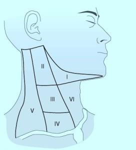
Neck Zones for Cancer

When thinking of neck zones, there are those zones used in trauma and there are those used for cancer surgery.

The neck zones or levels depicted here are for cancer surgery.  Lymph nodes or neck masses in these areas are described using these locations:

anatomical zones of neck
Picture copied from internet

  • In patients presenting with solid neck masses – those masses associated with the thyroid move with swallowing.  Those in levels 3,4, and 5 do not move with swallowing.
  • Metastatic lymph nodes found in level 6 – could come from thyroid.
  • U/S and FNA should be performed after exam/history for Solid Neck Mass.
  • With well differentiated thyroid cancers, there is a high rate of cervical metastasis particularly with papillary cancers.
  • With lateral lymph nodes involved in thyroid cancer, survival rate is not improved significantly with LN dissection in the neck for patients older than 60 years old or for men with a 4 cm or larger well differentiated cancer.
  • Prophylactic lymph node dissections does not seem to improve survival with patients undergoing thyroid cancer treatment.
  • If thyroid tumor is small with documented (U/S) lateral lymph nodes.  There is a benefit to dissecting level 3 or 4 lymph nodes (lymph node dissection or modified dissection) in the younger patient.

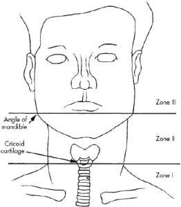
Trauma neck zones

picture copied from internet:
neck.zones


Newer Anticoagulants – Trauma teams having trouble reversing

Just recently a trauma nurse at Memorial Hospital, Modesto, CA was discussing with me the trouble with some of the newer anticoagulants.  She was saying that the bleeding was hard to reverse when the patients are on some of the newer anticoagulants.  I was trying to figure out which ones she was talking about and she said that if I had named them, then she would know.  So here’s my possible list of what was on her mind:

If it wasn’t Fondaparinux (above), then you’re probably thinking of Dabigatran/PRADAXA, Rivaroxaban/XARELTO or Apixaban/ELIQUIS. Can’t just push FFP, vitamin K or Factor VII for anticoagulation reversal.  Any one of these on your mind?

Trauma Pearls

  • 2 incision fasciotomy for compartment syndrome of extremity fractures – a common problem is incision is made too far laterally to help the anterior compartment (missed)
  • After 24-48 h of open abdomen management,  fistula rate is approx 15% if abdomen is not closed.
  • If stable hemothorax – 24-48 h to see if evac.  By day 2-3 it would be increasingly difficult to evacuate blood beyond that time, thorascopically.
  • Thoracic aortic injuries.  Endovascular repair typically not used in young patients or less than 20mm diameter aorta.
  • Complication of LMA = vomiting
  • Splenic injury, > or = 20% do not heal in three months.
  • Elderly patients with elevated INR, small head bleed on coumadin.  Rx with early plasma infusions, and early factor 7A use.
  • Carotid injuries do not occur commonly in easily accessible locations; put patient on antiplatelet  therapy and most of these injuries will heal.
  • Serial cardiac enzymes are rarely needed in patients with suspected blunt cardiac injury.
  • Zone I retroperitoneal hematomas = central periaorta hematomas.  Explore gun shot wound Zone I retrperitoneal hematomas even without expanding hematoma.
  • To evaluate  distal perfusion, proximity GSW to both extremities, use ankle brachial index rather than arterial pressure index.  (The API is the ratio between systolic blood pressures measured distal to a penetrating injury in one extremity and the systolic pressure measured at the same location in the contralateral uninjured extremity.)
  • solid organ injury, CT vascular extravasation should not determine management approach – instead depend on hemodynamics.
  • Injury to kidney after blunt trauma – prefer nonoperative treatment for better renal salvage even with urinary extravasation.
  • Head trauma, small epidural hematoma, no other injuries, observation in ICU x 24h – neuro exams, and repeat CT in 6h.
  • Leg with crush type wound with venous injury.  Re-establish blood flow quickly with shunt.  Tie a suture around shunt, do not debride the vessel back before placing shunt because the vessel will be injured more with the shunt tying.
  • Blunt traumatic arrest cases – not likely to survive to discharge from hospital.   Consider terminating resusitation.
  • Comatose patients, can remove cervical-collar if a good helical CT scan of the spine is completely normal.
  • A true transpelvic GSW (gunshot wound) should go to OR even with (or without) gross blood on rectal exam.
  • Complication and mortality rates  with rib fractures are twice higher in elderly patients than younger patients.
  • Physical exam with seatbelt sign is often not helpful, small bowel injury association is not 100%.
  • Side curtain airbags are more helpful than frontal airbags for additional protection in addition to seatbelts which do not do as well with lateral movement.
  • —-