Category Archives: Uncategorized

Hyperparathyroidism – primary, secondary, tertiary – simplified

The disease of hyperparathyroidism is diverse.  The causes of it are very different.  As a simplification there’s primary, secondary and tertiary hyperparathyroidism:

All three of these involves increased PTH (parathyroid hormone)

Primary: elevated calcium, low phosphorus in blood stream.  Usually caused by a parathyroid adenoma.

Secondary: normal or low calcium, usually caused by renal failure.

Tertiary: failure of treatment of secondary hyperparathyroidism to correct increased PTH.

More details on hyperparathyroidism

Primary hyperparathyroidism is most commonly caused by adenomas, greater than 85%.  Next most common cause would be hyperplasia which is 10% of the causes, and there is a 1% possibility that primary hyperparathyroidism is caused by a carcinoma.

Risk factor for primary hyperparathyroidism includes family history, MEN I and MEN IIa, and also radiation.  Major difference between MEN IIA and IIB:  MEN IIA has Hyperparathyroidism – hyperplasia, MEN IIB does not.  More on MEN syndromes.

Sestamibi scan is a method of detecting hyperparathyroidism.

Carcinomas which are commonly associated with hypercalcemia:

  • Breast Cancer Metastasis
  • Postate Cancer
  • Kidney Cancer
  • Lung Cancer

Another cause of hypercalcemia:  FHH – Familial hypocalciuric hypercalcemia

  • autosomal dominant
  • asymptomatic hypercalcemia and low urine calcium
  • with or without elevated PTH
  • No treatment is generally required for FHH
 

Primary hyperaldosteronism

Primary hyperaldosteronism

  • Hypertension in the presence of elevated aldosterone and suppressed plasma renin
  • Two major causes are aldosteronoma and idiopathic hyperaldosteronism (IHA)

Aldosteronoma

  • 60% of cases
  • more often in women
  • Hypertension and hypokalemia is more severe compared with IHA
  • Salt loading does not change aldosterone levels
  • CT usually reveals a small solitary adrenal tumor
  • Adrenal vein sampling will reveal unilateral hypersecretion of aldosterone.
  • Treatment is surgical resection

Idiopathic Hyperaldosteronism (IHA)

  • 30% of cases
  • More often in Men
  • Salt loading decreases aldosterone levels
  • CT reveals normal to bilaterally enlarged glads
  • Adrenal vein sampling reveals bilateral hypersecretion of aldosterone
  • Treatment is with potassium sparing diuretics – spironolactone


Somatostatinoma, Glucagonoma, insulinoma, vipoma, gastrinoma

Pancreas NeoEndocrine/Islet cell Tumors

Sometimes associated with MEN I (multiple endocrine neoplasia)

Somatostatinoma

  • tumor of the delta cells of the endocrine pancreas
  • triad of: Diabetes Mellitus, Steatorrhea, and Gallstones
  • Most are malignant and have metastasized

Glucagonoma

  • Necrolytic Migratory Erythema (NME).  found in 70%
  • Diabetes Mellitus
  • Most are malignant and have metastasized

VIPOMA

  • Vasoactive Intestinal Polypeptide tumor
  • Verner Morrison syndrome
  • Causes WDHHA:  Watery Diarrhea, Hypokalemia, Hypochlorhydria, Achlorhydria
  • Activation of cellular adenylate cyclase and cAMP production
  • 50% malignant

Insulinoma

  • 10% malignant
  • Whipple’s triad of pancreatic insulinoma – Hypoglycemia, symptoms of hypoglycemia, relief of symptoms when glucose is given.

Gastrinoma

  • Zollinger-Ellison Syndrome
  • Excess gastric acid production
  • Can arise in the duodenum or pancreas
  • 60% malignant


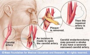
Indications for a Carotid Endarterectomy

When is a Carotid Endarterectomy Performed

Asymptomatic patients:

  • A Carotid Endarterectomy is indicated in patients with stenosis greater than 60%

Symptomatic patients:

  • A Carotid Endarterectomy is indicated in symptomatic patients with stenosis greater than 70%
  • If there are multiple episodes of TIAs, then operate when the stenosis is greater than 50%

carotidendarterectomy

Picture credit:

http://www.mayoclinic.com/health/medical/IM01657

Disclaimer: These are notes only.  Every patient and condition is different and requires a in person consultation with a medical doctor.  No medical advice is contained here, just academic discussion which may be very different from real life.   There’s always ongoing trials and the information here may be out of date.

Precedex does not cause respiratory depression

Dexmedetomidine (Precedex)

hp-precedexLogo

  • Sedation without respiratory depression
  • Ideal for use in the Intensive Care Unit.
  • Can be used for managing extubations.
  • No absolute contraindications
  • Compared to Versed (Midazolam), Precedex is associated with less delirium, tachycardia, and hypotension, but had more bradycardia
  • Agonist of alpha 2 adrenergic receptors in the brain
  • FDA reports that infusions greater than 24 hours in duration include ARDS, respiratory failure, and agitation


Trauma Pearls

  • 2 incision fasciotomy for compartment syndrome of extremity fractures – a common problem is incision is made too far laterally to help the anterior compartment (missed)
  • After 24-48 h of open abdomen management,  fistula rate is approx 15% if abdomen is not closed.
  • If stable hemothorax – 24-48 h to see if evac.  By day 2-3 it would be increasingly difficult to evacuate blood beyond that time, thorascopically.
  • Thoracic aortic injuries.  Endovascular repair typically not used in young patients or less than 20mm diameter aorta.
  • Complication of LMA = vomiting
  • Splenic injury, > or = 20% do not heal in three months.
  • Elderly patients with elevated INR, small head bleed on coumadin.  Rx with early plasma infusions, and early factor 7A use.
  • Carotid injuries do not occur commonly in easily accessible locations; put patient on antiplatelet  therapy and most of these injuries will heal.
  • Serial cardiac enzymes are rarely needed in patients with suspected blunt cardiac injury.
  • Zone I retroperitoneal hematomas = central periaorta hematomas.  Explore gun shot wound Zone I retrperitoneal hematomas even without expanding hematoma.
  • To evaluate  distal perfusion, proximity GSW to both extremities, use ankle brachial index rather than arterial pressure index.  (The API is the ratio between systolic blood pressures measured distal to a penetrating injury in one extremity and the systolic pressure measured at the same location in the contralateral uninjured extremity.)
  • solid organ injury, CT vascular extravasation should not determine management approach – instead depend on hemodynamics.
  • Injury to kidney after blunt trauma – prefer nonoperative treatment for better renal salvage even with urinary extravasation.
  • Head trauma, small epidural hematoma, no other injuries, observation in ICU x 24h – neuro exams, and repeat CT in 6h.
  • Leg with crush type wound with venous injury.  Re-establish blood flow quickly with shunt.  Tie a suture around shunt, do not debride the vessel back before placing shunt because the vessel will be injured more with the shunt tying.
  • Blunt traumatic arrest cases – not likely to survive to discharge from hospital.   Consider terminating resusitation.
  • Comatose patients, can remove cervical-collar if a good helical CT scan of the spine is completely normal.
  • A true transpelvic GSW (gunshot wound) should go to OR even with (or without) gross blood on rectal exam.
  • Complication and mortality rates  with rib fractures are twice higher in elderly patients than younger patients.
  • Physical exam with seatbelt sign is often not helpful, small bowel injury association is not 100%.
  • Side curtain airbags are more helpful than frontal airbags for additional protection in addition to seatbelts which do not do as well with lateral movement.
  • —-

Subclinical Cushing Syndrome

What is subclinical cushing’s syndrome?

  • subclinical hypercortisolism / may be observed with adrenal incidentaloma
  • autonomous clucocorticoid production without specific signs and symptoms of Cushing’s syndrome.
  • Much more common than classic Cushing’s syndrome
  • Patients have a high prevalence of obesity, hypertension, and type 2 diabetes.
  • Patients with incidentally detected adrenal masses who are about to undergo surgery should have testing for subclinical Cushing’s to avoid postoperative adrenal crisis..
  • Best test is short dexamethasone suppression test.

What is Cushing’s syndrome?

  • exaggerated facial roundness,
  • weight gain around the midsection and upper back
  • thinning of arms and legs.
  • stretch marks
  • hypertension
  • diabetes
  • Cushing’s syndrome occurs when exposed to high levels of the hormone cortisol for an extended period of time.
  • this can either occur with taking too much corticosteroid medication or when the body makes too much cortisol.
  1. Pituitary adenoma – secreting excess ACTH which stimulates the adrenal glands to make more cortisol
  2. Ectopic ACTH secreting tumor (ie. Lungs, pancreas, thyroid or thymus gland)
  3. Primary Adrenal Gland disease
  4. Familial Cushing syndrome

Diabetes type 1 vs. type 2

Type 1 = immune disorder.  Body attacks and destroys insulin producing beta cells in the pancreas.  Must take insulin.  Sometimes called insulin dependent or juvenile onset diabetes.

Type 2 = either the body doesn’t produce enough insulin or the cells ignore the insulin (resistant).  Sometimes called adult onset diabetes.  Obesity is the strongest risk factor for type 2 diabetes.

Adrenalectomy for subclinical Cushing’s Syndrome?

http://onlinelibrary.wiley.com/doi/10.1111/j.1365-2265.2011.04253.x/pdf

Adrenalectomy may lead to cure or better control of diseases associated with subclinical Cushing’s syndrome such as diabetes and hypertension

 Adrenal Incidentalomas

Most pose no clinical problems.

What is a Gastrointestinal Stromal Tumor (GIST)

GIST – Gastrointestinal Stromal Tumor

  • GISTs may be malignant or benign
  • More common in the stomach and small intestine but can be found anywhere along the GI tract.
  • Begins in cells called th interstitial cells of Cajal (ICC) – considered the pacemakers of the GI tract
  • GIST may have a genetic component.  Neurofibromatosis type 1 is linked to GIST.
  • Partial resections are usually adequate with GIST treatment with negative margins.  Formal gastrectomy is not usually necessary, nor is partial gastrectomy with nodal dissection.  Organ sparing approaches are usually appropriate.